

Date :
08/17/2026
Medically Reviewed By:
Dr. Samiullah Kundi, MD, Board-Certified Physician
Disclaimer:
The information provided on this page is for educational and informational purposes only and is not intended as a substitute for professional medical advice, diagnosis, or treatment. Always seek the advice of your physician or other qualified healthcare provider with any questions you may have regarding a medical condition or treatment plan.
Table of Contents
Pain in your back or neck is a brutal interruption. Suddenly, basic tasks require massive effort. In a strange twist, your own skeleton often causes this misery while trying to help. That biological backfire defines disc osteophyte disease. Cushions between your spinal bones wear out. Aging guarantees this. The body panics. It grows fresh bone spurs to lock up the wobbly joints. The catch? Those new, rigid bumps crush nearby nerves. Sharp, shooting agony follows. Fixing it starts with decoding exactly why it happens.
Professional Pain Recovery Support
If sharp, shooting pain from bone spurs is disrupting your daily routine, you don’t have to wait for the damage to worsen. At Indiana Neurology and Pain Center, our specialists can pinpoint the exact cause of your nerve compression and guide you toward a targeted, lasting solution.
The Mechanics Behind Disc Osteophyte Disease
Spinal mechanics explain the chaos. Think of the intervertebral discs resting between your vertebrae as tough rubber shock absorbers. Twisting, lifting, and simply standing upright rely entirely on them. They keep bones from grinding. Years pass. Miles add up. The discs dry out and flatten. Without that thick, hydrated padding, the spinal column wobbles.
Bones despise instability. To stop the dangerous shifting, your system deposits extra bone right on the vertebral edges. Doctors call this specific defense mechanism disc degeneration with osteophyte formation. Hearing “bone spur” makes people picture sharp, jagged spikes. Actually, these osteophytes are usually smooth and rounded. Their shape isn’t the issue. Location is everything.
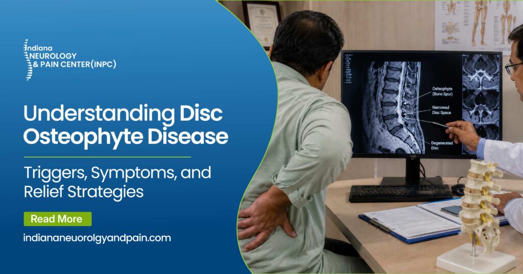
Merge a flattened, bulging disc with those invading bone spurs, and experts label the mess a disc osteophyte complex. When this mass shoves its way into the central spinal canal—or into the tiny side exits for nerves called neural foramina—the central nervous system takes a massive hit.
How Disc Osteophyte Disease Impacts the Nervous System
Most people assume the bone spurs themselves hurt. They don’t. Spurs lack nerve endings entirely. Pain only strikes when the osteophyte formation in spine joints begins scraping against nearby nerves, ligaments, or the spinal cord itself. Trapped nerves immediately fire distress signals. Your brain interprets those signals as sharp pain, numbness, or tingling.
| Structural Element | Healthy State | Degenerated State |
|---|---|---|
| Intervertebral Discs | Plump, flexible, and fully hydrated. | Flat, brittle, dry, and mechanically failing. |
| Nerve Openings (Foramina) | Wide channels that let nerve roots pass freely. | Severely narrowed by bone overgrowth, crushing the trapped nerves. |
| Overall Spinal Stability | Fully supported by elastic ligaments and thick, healthy discs. | Unstable, forcing the body to grow extra compensatory bone tissue. |
Identifying the Symptoms: From the Neck to the Lower Back
Symptoms depend heavily on the exact location of the spinal decay. Bone grows incredibly slowly. A person might not notice the structural damage for decades. The human body simply adapts until it runs out of extra space.
If the wear happens up high, it leads to a cervical disc osteophyte complex. The neck hauls around the heavy weight of the human head all day, making it a prime target for early joint failure. This issue triggers chronic stiffness and dull neck aches. But if a bony spur catches a cervical nerve root, pain shoots sharply down the shoulder, through the arm, and straight into the fingers. Patients often call it a sudden burning sensation or an electric shock. Hand weakness frequently follows if the nerve stays crushed.
When the lower back (lumbar spine) takes the hit, the symptoms shift downward. Sciatica dominates these cases. Sharp, shooting pain travels from the lower back deeply into the legs. Standing for hours makes lumbar symptoms worse. Sitting down and leaning forward, however, can sometimes pop the spinal canal open just enough to provide a brief window of relief.
Diagnosing Spinal Osteophytes and Disc Degeneration
Figuring out the exact pain trigger requires deep investigation. Medical teams spot spinal osteophytes and disc degeneration using physical checks paired with advanced scans. A normal clinic visit means testing reflexes, grading muscle power, and bending the spine to trigger that specific, familiar zap of pain. Finding that baseline tells the doctor which scans to order next.
- Standard X-Rays: Fantastic for looking at dense bone. Doctors easily spot shrunken spaces between bones and see the actual spurs poking into joint areas.
- MRI Scans (Magnetic Resonance Imaging): The top-tier choice for soft tissues. An MRI exposes exactly how dry the discs are and pinpoints exactly where hard bone is crushing a soft nerve root.
- CT Scans (Computed Tomography): These generate a sharp, cross-sectional 3D map of your skeleton. If a spine surgeon has to map out a precise, minimally invasive procedure, this scan is mandatory.
Identifying disc osteophyte disease early practically guarantees a wider menu of treatment choices—long before permanent nerve damage ruins the picture.
Effective Disc Degeneration With Osteophyte Formation Treatment
Calming angry nerves, lowering inflammation, and restoring normal movement sit at the top of the priority list. Surgery almost never happens right out of the gate. Doctors lean heavily into conservative care first to let the body heal.
Non-Surgical Treatment Approaches
The opening move for disc degeneration with osteophyte formation treatment zeroes in on intense rehab and crushing the pain immediately.
Physical therapy drives long-term success. Specific stretches and targeted lifting routines build up the core muscles wrapping your spine. Stronger muscles absorb the daily shock of walking and lifting. They function exactly like an internal back brace. Pressure comes right off the failing joints.
Medication handles the heavy lifting early in the process. Standard anti-inflammatory pills drain fluid away from trapped nerves. When pills fail, specialists usually switch to targeted steroid injections. Using live X-ray feeds, a doctor bathes the exact pinched nerve in potent drugs. Snapping that cycle of pain is vital. It finally allows a person to push hard in physical therapy without grimacing in agony.
| Approach | How It Actually Works | When Doctors Recommend It |
|---|---|---|
| Physical Therapy & Rehab | Builds up core strength to hold the spine upright and fix poor posture. | The very first step for nagging, moderate pain. |
| Epidural Steroid Injections | Floods the crushed nerve with strong anti-swelling drugs. | When sharp pain makes simple walking or exercising impossible. |
| Surgical Decompression | Cuts away the invading bone spur and any ruptured disc tissue. | After months of failed rehab, or if nerve damage is actively getting worse. |
Dealing with chronic nerve pain and spinal degeneration can drain you mentally and physically. If physical therapy, basic medications, and at-home care are no longer providing relief, our team is ready to step in. We offer advanced, minimally invasive treatments designed to relieve compressed nerves and get you moving comfortably again.
Advanced Osteophyte Disease Treatment
Sometimes the best rehab routines and perfectly placed injections just fail. Muscles might grow noticeably weaker. Numbness might slide further down a leg. Or the pain simply refuses to quit. That is when surgery becomes a necessary, highly effective osteophyte disease treatment.
Spinal decompression leads the surgical options. A specialized surgeon goes in to manually shave down the invading bone spurs and the thick, overgrown ligaments choking the nerves. If a joint is severely destroyed, they might remove the ruined disc entirely and fuse two adjacent bones together. A fusion permanently halts the agonizing bone-on-bone friction. Thanks to better tools, surgeons handle many of these fixes through tiny incisions. Less muscle cutting means minimal bleeding and a radically faster recovery.
Dealing with disc osteophyte disease drains you mentally and physically. However, the vast majority of people eventually lock down real, lasting relief by following these exact clinical steps.
Take the Next Step towards Better Help

Dr. Samiullah Kundi
Pain medicine & Neurologist
Dr Kundi is a board-certified neurologist with rigorous medical training and pain management expertise. Mr. Kundi has been certified by the American Board of Pain Medicine (ABPM), American Board of Psychiatry and Neurology (ABPN) – Clinical Neurophysiology American Board of Integrative Holistic Medicine (ABIHM), and American Board of Psychiatry and Neurology (ABPN) – Neurology. Dr. Kundi’s vision of serving people with neurological pain has led to the establishment of the Indiana Neurology and Pain Management Centre.
Frequently Asked Questions
No. Solid bone never just melts or shrinks away. But here is the bright side. You rarely need to remove the spur to feel better. Killing the surrounding inflammation usually makes the pain vanish entirely.
Not exactly. Thousands of older adults walk around with massive bone spurs and completely flat discs while feeling perfectly fine. Agony only strikes when that extra bone physically crashes into a nerve or hits the spinal cord.
Smoking wrecks your spine. Nicotine cuts off blood flow, starving your discs of essential nutrients. Carrying heavy excess body weight also accelerates the breakdown by forcing the spine to haul an extreme mechanical load every single day.
No. A herniated disc happens when the soft, jelly-like inside squishes out of its casing. A bone spur is hard, rigid bone. They are different, yet they frequently show up in the exact same spot, building a tangled, painful mess.
It hinges entirely on the exact operation. A minimally invasive microdiscectomy could have someone sitting back at a desk job in a few weeks. A major spinal fusion? That demands months of cautious rehab before attempting heavy lifting.